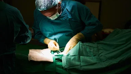
There are risks inherent in the procedure. In the case of full thickness burns, in which both the epidermis and dermis have been destroyed, there may not be enough healthy skin available to cover the burn. The harvesting of healthy skin can also result in severe blood loss, though there are methods that can reduce the extent of it. Either way, healing of both the donor site and the burn site can take weeks, and long-term problems for the patient include scarring, decreased sensitivity and infections. Could there be a better way? Jeschke and colleague Axel Guenther, an associate professor in mechanical engineering at the University of Toronto, think so. They believe they have found an alternative to autologous skin grafts that could revolutionise the treatment of burns patients.
Biosynthetic alternatives are timeconsuming and costly
Researchers have looked around for alternatives to skin grafts for some time, such as using biosynthetic products that combine bovine collagen, synthetic materials and human donor skin. The “gold standard” technology, says Guenther, is based on collagen, which promotes healing, and is the main protein constituent of intact skin. This technology, developed more than 30 years ago, is expensive, however, and in many healthcare systems is predominantly used for injuries to the face and hands. In the past ten to 15 years, Guenther adds, a number of cell-based solutions have been developed, but they are also “extremely expensive” and “require a lot of time”. It’s the advent of 3D printing that has enabled Guenther and Jeschke to take a dramatic step forward in the treatment of burns. In conventional 3D printing, or additive manufacturing, a model of the desired object is created using computer-aided design (CAD) software. This contains instructions that are then sent to the 3D printer, which extrudes a layer of material (plastic or powders, for example) through a nozzle to create a single slice of the object. As that cools, the printer then continues depositing layers until a solid object is created.
In the past ten to 15 years, we’ve seen increasing scientific interest in 3D bioprinting. This uses the same technique, but this time the object created is a human organ, and the materials deposited are a combination of human cells and hydrogel-based scaffolding materials to sustain them. This combination of cells and hydrogel-based materials is known as bio-ink.
The technology has obvious application in the area of burns. 3D bioprinters enable skin cells, along with the scaffolding materials, to be deposited layer by layer over the burn. “There’s a lot of interest in using general printing technologies, particularly extrusionbased printing technologies, in burn treatment,” Guenther explains, “and lots of the alternative systems that have been recommended are rather conventional 3D printers scaled to the patient size. So you have a printer that moves in all three directions, which is in principle a viable approach.”
Such systems are, however, large and costly. While they may work in the handful of places that can afford them, Guenther and Jeschke concluded a more scalable system was needed. This scalable system could, explains Guenther, “be used in any kind of non-super-specialised setting but also be used potentially in very hard-to-reach places outside of
operating rooms”.
A 3D printer that works like a paint roller
Over the course of eight years, the pair have collaborated to develop a portable, handheld 3D bioink printer that can replace the traditional skin graft. It works a little like a paint roller, depositing a sheet of biomaterial on to the wound. “It’s like a duct tape dispenser that you buy in the hardware store, with the difference that there is not a roll of tape that you unroll, but there is a print head that squishes out the soft layer of, so to speak, skin tissue tape lightly on to the wound surface,” says Guenther, adding, “I think it has the potential to become a game changer.”
The bio-ink contains a biopolymer solution made out of fibrin and collagen, the proteins involved in wound healing. This biopolymer is converted into a gel through a cross-linking mechanism involving a second fluid, thrombin. The cells used in the bio-ink are stem cells from the burn site – the discovery made by Jeschke’s team two or three years ago that burnt skin contains viable stem cells was hugely significant in the development of the technology.
“Even though the tissue may be severely injured, it does still contain stem cells that, if isolated and reintroduced in the appropriate matrix, can promote wound healing, which is quite remarkable,” says Guenther.
There have been multiple challenges in developing a workable product. The skin is a complex organ containing different types of cells, so converting the stem cells into the desired skin cells is, as Jeschke puts it, “not very trivial”. Another difficulty is in applying the solution to the burn site. “If you wanted to apply a homogenous layer of the biopolymer on top of the wound, it’s not an easy undertaking, because it’s quite a liquid solution, so it would drip off a wound,” says Guenther. “Wound surfaces, unlike dishes in our cell culture facilities, are never flat, and nor are they horizontal. The standard wound is curved, usually with a pretty high inclination angle, so the homogeneous deposition of the precursor ink and the conversion into the gelled material isn’t trivial.”
The way he and Jeschke solved this, he explains, is by using microfabrication technologies. A chip with a large number of parallel panels squeezes out identical measures of the biopolymer solution, and a second array of similar channels across the top squeezes out the cross-linker. “And so as that chip homogeneously travels over the topology of the wound, it leaves behind a very consistent layer of a rapidly cross-linked precursor bio-ink that gets transferred to precursor tissue,” Guenther explains. “And that’s the basic innovation behind the approach.”
Human trials within two years
The pair revealed their first prototype in a paper published in 2018. Since then, there have been at least ten more, as they work towards creating a product that can be used by clinicians. But how near are they to a commercial version?
Currently Jeschke’s laboratory at the Sunnybrook Research Institute is carrying out a large animal pre-clinical study, using the printer to treat burn wounds on pigs. Human trials will follow in the next two years.
The animal studies have been successful in demonstrating that the approach accelerates wound healing, says Guenther, but that’s only the first step. “Current work is directed towards improving the composition of the bio-ink in the sense that we don’t only accelerate wound healing, but ideally also reduce the amount of scarring that goes along with wound healing,” he adds. “So priority one is that the wound is healing rapidly, but it does make a difference to the patients’ lives whether the wound is highly scarred or the amount of scarring is significantly reduced.”
There is still much to be done before the product reaches the market. Work on redesigning the core parts of the printer, so that it can be scaled up and mass produced, will begin this autumn. “The biggest bottleneck,” says Guenther, “is the cells, which is the case for any cell therapy. Making patient cells available locally, at large quantities and low cost is challenging, but there is dramatic progress being made over the last years.” Regulatory approval will also have to be sought.
Axel Guenther
Once the product comes to market, Jeschke believes that the more rapid healing and the lack of scarring will “really move patient care forward”. And although the initial work is being done on burns, the principle can, he says, be applied to any wound: “We just have the burn patients because that is my patient population, but my vision is to make this more broadly available and to go even further than just burn patients.”
The team is confident that its meticulous approach will reap benefits. “Part of the reason why it’s taken seven years is that we have reproduced every single data set along the way,” says Guenther. “We want to really make sure that the things that leave the lab and are moving into the commercialisation realm are rock solid from a scientific perspective.”
Reducing scarring
Guenther and Jeschke’s isn’t the only long-term Canadian project focusing on helping burns heal and reducing the scarring that accompanies the process. Led by Jeff Biernaskie, professor of stem cell biology and the Calgary Firefighters Burn Treatment Society Chair in Skin Regeneration and Wound Healing, researchers at the University of Calgary have identified what separates the dermal progenitor cells that are able to regenerate new skin and those that create disfiguring scar tissue. “Remarkably, we found that although these cells come from the same cellular origin, different microenvironments within the wound activate entirely different sets of genes,” explained Biernaskie. “Meaning, the signals found within ‘regenerative zones’ of the wound promote reactivation of genes that are typically engaged during skin development; whereas, in scar-forming zones these pro-regenerative programmes are absent or suppressed and scar-forming programmes dominate.” That discovery looks like it could be the foundation for a number of exciting, life-changing developments. The research team has also shown that it’s possible to modify the genetic programmes using artificial stimuli. “What we’ve shown is that you can alter the wound environment with drugs, or modify the genetics of these progenitor cells directly, and both are sufficient to change their behaviour during wound healing,” Biernaskie continued. “And that can have really quite impressive effects on healing that includes regeneration of new hair follicles, glands and fat within the wounded skin.” With proof of principle that adult wound-responsive cells have a latent regenerative capacity, the Calgary research team is exploring other pathways involved in drawing it out. Their eventual hope is to develop a cocktail of drugs that can support wound healing by preventing the genetic programmes that lead to the formation of scar tissue and encouraging progenitor cells’ regenerative capacity. Five years in the making, the latest study was published in the scientific journal Cell Stem Cell in August.


