Technological shortfalls in medical imaging have consistently been a problem in medicine; as soon as something is developed, the team behind it will start working on something else. Radiologists and urologists have been frustrated for decades by the inability of conventional imaging tests – including CT and MRI scans – to distinguish between benign and malignant kidney tumours.
In the US, 16 out of 100,000 men and women are affected by kidney and renal pelvis cancer every year. Out of the 13,000 new kidney cancer diagnoses annually in the UK, 4,000 result in loss of life. According to Cancer Research UK, there is a 50% chance of survival. Tumours in the kidney can be benign or malignant; the former is not as harmful and can take the form of cysts.
Dr Mehrbod Javadi, director of nuclear medicine, and assistant professor of radiology and radiological science, formed part of a team of scientists – which also included Mohammad Allaf, vice-chairman of urology – at the prestigious Johns Hopkins Medicine in Baltimore, US, to conduct research on designing studies and performing analysis in relation to developing new techniques for non-invasive imaging tests, which will be more accurate in ruling out specific kidney cancers. It was also hoped that this would pave the way for establishing new methods to characterise renal masses.
Javadi discusses these studies and how they boost the accuracy of kidney tumour classification, as well as the addition of a 99mTc-sestamibi SPECT/ CT test to CT or MRI scans.
In early 2017, the research team reported that the potential improvement in diagnostic accuracy will prevent painful surgical intervention for thousands of patients across the US.
Andrew Putwain: Can you tell us more about how the technology behind the test works, and what its capabilities are beyond imaging technology?
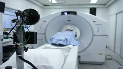
Mehrbod Javadi: We use a radio -labelled molecule called 99mTcsestamibi, which crosses into cells and binds active mitochondria because of the large negative potential across the mitochondrial membrane. We then use a high-resolution hybrid gamma camera and CT systems to image where the sestamibi concentrates in tissue, and fuse them to CT images to create a molecular and anatomic hybrid map of the kidneys. This map tells us if the renal tumours concentrate the sestamibi molecule or not.
Tumours that have high mitochondrial concentrations, such as benign oncocytomas, concentrate the radioactive tracer, whereas malignant tumours – including renal cell carcinomas (RCC) – do not concentrate the sestamibi. This hybrid approach of using the sestamibi mitochondrial maps and anatomic CT imaging combines two different, yet complementary technologies, providing anatomic and molecular/functional data in one test.
Anatomic-only studies, such as IV contrast CT or MRI, are unable to effectively distinguish between renal oncocytomas – which are the most prevalent benign tumours of the kidney – and clear cell renal cell carcinoma (CCRCC), a common malignant tumour of the kidney.
The publicity for the study outlines how it can prevent more invasive treatments being undertaken. Can you explain why this is the case and how it will benefit patients?
By having a non-invasive imaging test, with a diagnostic performance that is similar to or better than a biopsy, patients can be risk stratified into low and highrisk groups. More than 5,000 patients with benign oncocytomas have their kidneys resected every year in the US. The study suggests that there may be a non-invasive path for doctors to take that characterises benign tumours without surgery.
Conventional procedures for renal tumour imaging include multiphase IV contrast-enhanced CT, MRI and ultrasound. None of these techniques can reliably distinguish the most common benign renal tumours – oncocytomas – from prevalent malignant renal tumours, CCRCC. As a result, the majority of these patients undergo an invasive biopsy or surgical resection of the lesion. Unfortunately, a biopsy can create complications and can be non-diagnostic or under -sampled in up to 20% of patients. In addition to its morbidity and mortality, surgery often results in removing the entire kidney or part of it.
Why did your team decide to work on this piece of technology and what was the end goal – a more advanced imaging system or curtailing the number of invasive treatments?
As the use of conventional crosssectional imaging has increased, radiologists are detecting more renal tumours. These tumours have been a clinical conundrum for urologists, as 15–20% of the tumours resected are benign lesions that did not require surgical intervention.
My colleagues, Dr Michael Gorin and Dr Steven Rowe, assistant professors of urology, and radiology and radiological science, respectively, hypothesised that applying modern high-resolution hybrid SPECT/CT technology to a technique that saw Dr Thomas Gormley, and his colleagues, outline that benign oncocytomas could be separated from malignant RCC, helping to decrease the number of unneeded kidney resections for benign lesions. Gormley, a urologist from Rio Grande Urology, Texas, published this data in a 1996 study called ‘Renal oncocytoma: preoperative diagnosis using technetium 99m sestamibi imaging’.
Why are treatments so hard to administer to the kidney and what makes its tumours difficult to classify?
The treatment for kidney tumours is surgical resection. Even with excellent technique and experience, there is a risk of infection in surgery, bleeding and post-operative complications.
The difficulty in classification by imaging stems from the similar anatomic characteristics of oncocytomas and CCRCCs. From a vascular and macrostructural standpoint, these tumours appear to be very similar. The difficulty in histopathological classification generally lies in sampling errors, which are often from small tumours and can be heterogeneous. The under-sampling of a tumour may result in incomplete histopathologic characterisation.
What cons could be taken from a more advanced imaging system?
Mainly imaging time: the procedure requires that the patient be on the scanner for approximately 30 minutes, and then they are in the molecular imaging department for up to two hours because of the uptake time required.
What do you hope further studies will discover about 99mTc-sestamibi SPECT/CT?
I think there are a few areas where we need to learn more. Firstly, we need to optimise the imaging procedure itself, and find out what the optimal imaging time points and optimal imaging reconstruction algorithms are, and whether or not there are quantitative tools that can help doctors with characterisation.
Also, and perhaps more importantly, we need to expand the research and imaging by studying more patients. Our initial imaging trial has been confirmed by a separate non-affiliated group from Sweden, but I think having more patient data would enable us to make more concrete risk assessments.
And thirdly, I think we need to take this imaging framework, together with larger patient data sets, and work beside the surgical and imaging communities to develop diagnostic algorithms that can help to improve how we risk stratify individuals, in order to save patients with low-risk tumours from having unnecessary surgery.
Where do you see this type of technology going?
Sestamibi is an established, inexpensive and readily available radiotracer that is typically used in myocardial perfusion and parathyroid adenoma imaging. The combination of this imaging tracer and a renal carcinoma-specific imaging tracer could help to enhance the performance of hybrid SPECT/CT imaging of the kidneys.
Furthermore, renal tumour imaging is a very promising application for quantitative SPECT/CT. Quantitation allows the use of multiple imaging biomarkers, some of which will likely help in assessing renal masses with molecular imaging techniques.
The future in spec
“Although further study is needed to validate the accuracy of 99mTc -sestamibi SPECT/CT, this test appears to be a less expensive, faster and non-invasive alternative to surgery,” adds Gorin, who is also chief resident at the James Buchanan Brady Urological Institute at Johns Hopkins University School of Medicine. “In the absence of diagnostic certainty, surgeons tend to remove kidney tumours in an abundance of caution, leading to an estimated 5,600 surgically removed benign kidney tumours each year in the US.” With those sorts of numbers, it is not hard to see why a new procedure needs to be introduced.


