Americans love their football, and they get started early: high school games can draw enormous crowds. But American football, or gridiron, is a high-contact, sometimes violent sport where athletes are required to wear heavy protective gear. Many professional games results in at least one concussion. The size of the players doesn’t seem to correlate with the head injury rates: high school players face double the concussion risk compared with college players, who are the next level down from the pros. While the National Football League (NFL) has put several measures in place to reduce concussion rates for adult players, a growing body of research suggests playing gridiron at a young age carries a high possibility of inflicting permanent brain damage.
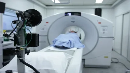
With those figures in the back of their minds, and no clear guidelines on how to proceed with a young patient with minor head trauma, physicians and parents tend to opt for a computed tomography (CT) scan just to be safe. Rates of CT head scans have been rising steadily in the US, from three million a year in 1980 to 80 million in 2016, and the vast majority of CT head scans for minor injuries show no signs of traumatic brain injury. US FDA estimates that somewhere between 10–30% of CT scans on children are medically unnecessary. CT remains an essential diagnostic tool, but medically unwarranted paediatric head scans may be doing more harm than good.
Multiple scans, more risk
A CT scan delivers a dose of radiation roughly 200 times as high as a chest X-ray. Multiple scans can quickly add up to a dangerous amount of radiation exposure, which can mean an increased cancer risk later in life. There have been a number of studies into exactly how high that risk might be; one 2009 study by the US National Cancer Institute, led by Amy Berrington de Gonzalez, estimated that the 72 million CT scans performed in 2007 would translate into 29,000 future cancers and 14,500 related deaths.
Children are more susceptible to the effects of radiation, so another study, published in 2012 and also led by Berrington de Gonzalez, looked at 180,000 British patients who had been scanned before they turned 22. It found that having two to three head CT scans before the age of 15 tripled a patient’s risk of brain cancer; five to ten scans would triple the risk of leukaemia. So, while one scan on its own has a negligible risk, the danger starts to become very real for patients like highschool footballers, who might be scanned multiple times over their athletic careers. The problem for physicians is that they often don’t know whether a scan was necessary until after they’ve performed it.
In New Jersey, 20% of boys who play sports in high school are football players, making it the leading high school sport by a wide margin. That adds up to a lot of bumps on the head during practices and games, and an accompanying number of ‘just in case’ CT scans. In mid-2016, a collective of New Jersey medical associations announced a drive to cut down on unnecessary paediatric head scans throughout the state. The New Jersey Hospital Association’s Institute for Quality and Patient Safety partnered with the Hospital Improvement Innovation Network and the New Jersey Council of Children’s Hospitals to create the Safe CT Imaging Collaborative, along with 47 hospitals. They gathered data from New Jersey’s 71 acute-care hospitals to measure rates of diagnostic CT scans for head trauma, and then teamed up to send clear educational materials to every hospital in the state on when and how to scan responsibly. After a year, they’ve decreased CT scans on minor head injuries across the state by 25%, or almost 1,000 scans.
New guidelines
Dr Ernest Leva is the physician chair of the NJ Council of Children’s Hospitals, as well as an associate professor and director of the Division of Paediatric Emergency Medicine at the Robert Wood Johnson Medical School at Rutgers University. He says that, up until now, one of the major drivers for unnecessary CT scanning has been a knowledge gap in hospitals.
“The body that formulated those guidelines is a paediatric body,” Leva explains. “Most children are seen in emergency departments that are staffed by emergency medicine doctors. Generally, if it’s not your organisation, you get people who don’t necessarily abide by it or even are aware that it exists.”
The group he’s referring to is the Paediatric Emergency Care Applied Research Network (PECARN), the first federally funded network of its kind in the US. The Safe CT Imaging Collaborative’s guidelines are based on a PECARN study published in 2009 and updated in 2015.
The PECARN rules are not the first to cover CT scanning in children. Previous sets of guidelines have included the 2006 Children’s Head Injury Algorithm for the Prediction of Important Clinical Events and the Canadian Assessment of Tomography for Childhood Head Injury, which used large sample sizes. But the PECARN study was bigger, and the prediction rule it came up with turned out to be more accurate and encompassed more risk areas.
The study wasn’t aimed at predicting cancer risk or identifying patients in danger of brain damage. Instead, its goal was to find out which children were at “very low risk” of traumatic brain injury after minor blunt trauma to the head. The researchers wanted to give doctors more confidence when deciding to place a patient under observation instead of scanning.
Rules of engagement
To come up with the prediction rule, the researchers looked at cases involving more than 42,000 children presenting with minor blunt trauma to the head at hospitals across the US. Overall, just over 5% of the patients ended up with a clinically important traumatic brain injury (ciTBI). They broke down the patient group by age and scores on the Glasgow Coma Scale (GCS) – an assessment that measures eye opening, and verbal and motor responses to decide on a patient’s conscious state. A score of 15 means the patient is fully awake, while a score of three or less indicates the patient is either dead or in a deep coma.
Broadly, the PECARN rules recommend a CT scan if the patient scores a GCS of 14 or shows signs of a skull fracture – this group had up to a 4.4% risk of ciTBI. It’s up to the physician to decide whether or not to scan if patients don’t meet those criteria, but do have a bruise, a bad headache, or a history of losing consciousness or vomiting; if they were hurt in a severe way such as a car accident; or if an infant’s parent say the child is acting strangely. The rules differ slightly for children under and over two years old, but when none of those signs are present in either age group, there is less than a 0.05% risk that the patient has ciTBI, so it’s better to keep an eye on them instead of scanning.
“I think [the PECARN rule] is a good tool to use,” says Leva. “People know radiation is dangerous, but I don’t think they understand that there are real, good guidelines that they can follow to decrease that risk.”
The parent trap
But even when physicians are au fait with the latest research, parents with no clear information about cancer risks may request a scan for peace of mind in the moment. For the scan-reduction campaign to be successful in the long term, the general public needed to properly understand the risks.
In September, the New Jersey collaborative launched the #ScanSmart initiative, aimed at parents, trainers and high-school athletic coaches. They distilled the guidelines into an easy-to-remember acronym, which they called the COOL approach: consider using other kinds of testing when appropriate; only image the indicated area; only scan once; lowest amount of radiation, based on child dosing.
Leva says parental pressure is “definitely a driver” of the rising rates of CT scans. The collaborative hasn’t put together any guidance for physicians on how to convince a parent that it’s better for their child not to receive a CT scan. Rather, the hope is that #ScanSmart and the COOL system will do the work of educating parents. Once they’re aware of the need to weigh the risk of cancer against the danger of brain damage, they can self-advocate.
“In one of the educational packets, we also have a little list where parents can write how many times their kids have had CT scans,” Leva says, adding that the community has been “very receptive”.
Pressure to scan can, of course, come from the opposite direction: it’s the easier option from an operational point of view. “It’s more labour-intensive, because the patients are around more, so they have to be observed more,” Leva explains. “From a cost-benefit ratio, the patients are not charged for the CT scan or the interpretation by the radiologist so it’s a large saving [for the patient], but there is a little bit more human capital involved.”
Losing the revenue of nearly 1,000 scans a year in favour of spending more staff time observing patients may not sound like a profitable trade-off for hospitals, but Leva says he’s had “no pushback at all” on that subject from New Jersey’s healthcare system. “I think people are very willing to do that in order to do a better job,” he says.
Safe scans
The Safe CT Imaging Collaborative plans to reduce head CT scans in New Jersey by another 25% this year, as well as launching a second initiative to combat CT scan rates for abdominal pain.
“It’s much more difficult with abdominal pain, because there’s no good data, like you have with the PECARN initiative and minor head injury, but we are looking to push for more ultrasound and MRI evaluation,” Leva says.
It’s easy to take a ‘better safe than sorry’ approach to scanning in paediatrics; no one wants to risk allowing a child with brain damage to go untreated. But in a community like the US high-school sports system, where minor paediatric head traumas happen regularly, everyone needs to understand how to walk the line between feeling confident in the moment and staying safe in the long term. Clear guidelines and a sustained public campaign can take out the guesswork and leave doctors, patients and parents reassured.


