Throughout most of history, surgery meant one thing only: opening up the patient’s body and directly working on the surgical area. Whether it was an appendectomy or a gynaecological procedure, the patient faced what could sometimes be a traumatic and risky operation. Although the idea of minimally invasive surgery had been touted for some time, it was only in the early 1980s that the vision became a reality.
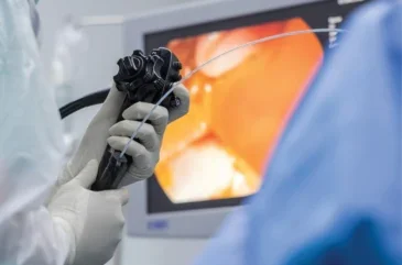
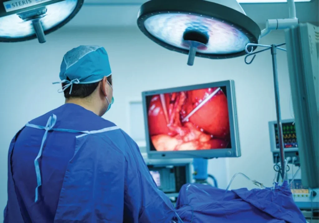
Since then, the field of minimally invasive surgery has been fast advancing. The first technique to arrive on the scene was laparoscopic, or ‘keyhole’, surgery, which involves making small incisions in the abdomen or pelvis and inserting a thin tube with a video camera. If necessary, additional surgical instruments can pass through the incisions too, meaning surgeons can perform intricate procedures guided by a video display.
From the patient’s perspective, keyhole surgery is associated with less pain, shorter hospital stays, quicker recovery times and fewer complications. There are benefits for health systems too.
One study found that as laparoscopic surgeries for colon cancer increased, the costs for the NHS fell dramatically – between 2006 and 2012, this new approach saved £29.3m.
All that said, new techniques come with new challenges. As Professor Kaspar Althoefer, an electrical engineer and roboticist at Queen Mary University of London points out, the surgical instruments themselves need to be miniaturised so they can fit through the incisions. Meanwhile, the surgeon no longer holds the tool directly – he or she clutches the end of a long, thin laparoscopic device, which enters the patient’s body via a trocar port.
“That is very challenging,” says Althoefer. “You have these laparoscopic tools going through these narrow ports, and the surgeon can move them in one direction. But because there is a pivot point at the trocar port, any movement in one direction will lead to the opposite movement at the other end. That’s very complicated, especially for something like suturing, and surgeons may need many years to perfect it.”
For this reason, many surgeons have gravitated towards robot-assisted minimally invasive surgery (RAMIS), which can perform motions beyond the scope of a human hand. Rather than holding the laparoscopic devices manually, surgeons use an input device resembling a joystick, enabling them to move these tiny instruments with incredible precision.
The dominant market player here is the da Vinci Surgical System from Intuitive Surgical, which has been used across 10 million procedures (especially urological surgeries) since it was launched at the turn of the millennium.
“The devices are now controlled by a robot arm, and one can write clever algorithms to make it much easier to control what’s happening inside the patient,” says Althoefer. “Suturing becomes very easy – I was able to make a knot in two minutes in a test environment, and I’m an engineer.”
Softly does it
Althoefer describes these techniques as a “great step forward”, remarking that RAMIS in particular is a “tremendous development”. However, he feels that there is still room for improvement. For the past 12 years, his work has focused on soft robotics for minimally invasive surgery – soft robot manipulators made up of biocompatible silicone rubber with inflatable chambers inside.
“In 2012, we started an EU-funded large-scale project called STIFF-FLOP,” says Althoefer. “The idea was to move radically away from these rigid tools and to create robots that are made from soft materials. Since the device is soft, operating inside a human becomes much safer.”
Although systems such as Da Vinci do have inbuilt safety mechanisms, the rigidity of the materials can occasionally lead to patient injury. This is especially true in the case of software hiccups, which might cause the robot to move in undesired ways. Soft robotics would minimise these risks while vastly improving flexibility.
“Our systems can be likened to the arms of an octopus,” says Althoefer. “They are very soft, but at the same time, can move in very dextrous ways into spaces like the abdominal cavity, moving easily around organs to reach the area of interest.”
Although there is still work to be done in terms of precision and accuracy, Althoefer believes soft robotics could hold great potential over the years to come. His team are starting a new project, funded by the European Research Council, to develop a ‘next level colonoscopy’ – a soft robotic device that would travel along the colon to carry out both diagnostics and surgery.
The device in question, known as an ‘eversion robot’, contains a sleeve that grows out from the tip when pressurised, enabling the robots to move forward in a frictionless way. This would greatly improve comfort levels for the patient. “We hope our new approach would convince more people to have this examination done, so that we can effectively save more lives in the long run,” says Althoefer.

Another team, from the Massachusetts Institute of Technology, have been working on a different solution to this problem. Frequently, patients undergoing a colonoscopy will need to have polyps removed – a situation that can sometimes lead to gastrointestinal bleeding. The MIT researchers have designed a sprayable gel, GastroShield, that could be applied onto the surgical sites through an endoscope. The gel forms a protective layer, preventing bleeding during and after the procedure.
The role of AI
While some researchers are focused on patient comfort levels, others are more interested in finetuning the information the surgeon receives on their screen. One example is EnAcuity, a spin-out company from University College London and Imperial College London. They are developing an AI-based technology that can be integrated with existing laparoscopic cameras, enhancing the visual output.
“Our product offers a breakthrough solution in AI-driven tissue oxygenation monitoring during minimally invasive surgery,” is how Dr Maria Leiloglou, CEO of EnAcuity, puts it. “Hyperspectral Imaging (HSI) allows for the capture of small differences in colour. This information is analysed to calculate the oxygenated fraction of blood in the tissue. EnAcuity’s solution predicts HSI from standard laparoscopic images, providing real-time insights into tissue perfusion. This is crucial for reducing severe surgery complications.”
There are various other solutions emerging in this space, typically requiring specialised hardware. However, because EnAcuity’s technology is software-only, it keeps costs to a minimum; hospitals won’t need to invest in additional cameras or spend lots of time on training or maintenance.
Having successfully concluded proof-of-concept studies, the developers are now commencing video data collection at collaborating hospitals, which will help them fine-tune their software ahead of launch. “EnAcuity’s aim for this year is to tailor and validate the use of the software for real-time tissue oxygenation with the use of human colorectal and gallbladder video data,” says Leiloglou, who is also an honorary research fellow at UCL and Imperial. “Following this milestone, our seed-round raise will commence, enabling the product’s release into the UK market.”
EnAcuity’s technology is just one example of the ways AI is being used in this space. As Leiloglou explains, minimally invasive surgery is constantly improving, reducing the risk of patient complications with every new technological breakthrough. AI will likely accelerate this trend, especially when it comes to intraoperative imaging, but also in relation to factors like surgeon workflow and equipment use.
Althoefer thinks human surgeons shouldn’t hang up their gowns just yet; we are still a long way from a surgical system that doesn’t require any human interference. That said, he agrees that the field is progressing quickly. He gives the example of tremor reduction, which is being used within the da Vinci system already; you can programme your robot not to react to bodily tremors, giving rise to a steadier movement.
“The hope is that AI could possibly take over more and more from the surgeon,” he says. “For example, routines could be learned within an AI framework to carry out a suturing task. The surgeon would just identify the start point and the end point and then the robot would automatically do the suturing across a section of tissue.”
Other advances and developments
Another intriguing development, already being used within robotic systems, is one that could bring surgery full circle. “If you go back to the very beginning of open surgery, where the surgeon would put their hands inside the belly, they would get haptic feedback,” says Althoefer. “They could even close their eyes and understand the anatomy, making it easier to make decisions. Using laparoscopic surgery, or even worse, RAMIS, there is no more touch feedback, and that is quite limiting.”
Newer approaches aim to restore this haptic feedback, by measuring the way the robot tip interacts with the environment and feeding that information back to the surgeon. A 2023 research review found that haptic feedback typically reduced the forces applied during surgery, as well as reducing the completion time, improving accuracy and enhancing success rates.
In terms of what else may be coming down the line, Althoefer mentions a new type of procedure called natural orifice transluminal endoscopic surgery (NOTES), which involves a flexible endoscope that can be inserted through an existing orifice like the mouth. From there, it can travel into the stomach via the oesophagus, and from there into the abdominal cavity where the operation takes place. While most of the work in this space is a long way from the clinical market, it could be very promising over the longer term.
Similarly exciting, though similarly speculative, are micro-robotic devices, which could notionally travel through blood vessels and assemble inside the body to carry out a task. As Althoefer puts it: “Moving from laparoscopic surgery mainly in the abdominal area, to using a robot was already an enormous step. But there are many new things happening in this domain.”


